How the Heart Shapes the Mind: A new approach to emotion
Imagine that you are driving your car when, suddenly, a large rock blocks your way. Shocked, you applied the brakes. Now, consider how we traditionally understand brain function through this example: the brain detects the sudden danger and triggers a bodily response, causing you to hit the brake pedal.

However, according to the studies conducted by Brian Hsueh, Ritchie Chen, Karl Deisseroth, and their colleagues at Stanford University proposes a fundamentally different perspective. To understand this idea, imagine that you are driving along a clear road when a red warning light suddenly starts flashing on your dashboard. You see no boulder or other obvious danger, yet the flashing light makes you nervous. Your mind immediately begins to interpret the signal as evidence that something is wrong. In this analogy, the dashboard warning light represents your heartbeat.
Methodology and Experimental Models
To investigate this bottom-up flow of information from the body to the brain, the researchers used mouse models, including wild-type C57BL/6J mice and transgenic TRAP2;Ai14 mice.
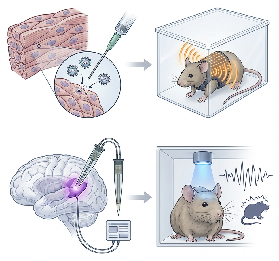
Until recently, manipulating heart rate in awake animals without causing pain, tissue damage, or unwanted drug effects had been technically challenging. To overcome this limitation, the researchers developed a noninvasive optical pacemaker.
First, they administered an AAV9 viral vector through a systemic retro-orbital injection. Using the cardiac troponin T promoter (mTNT), they targeted the expression of the light-sensitive protein ChRmine to cardiomyocytes, the muscle cells responsible for heart contraction. This approach was designed to minimize unwanted expression in neurons and other organs.
Next, the team developed a wearable fabric vest containing a micro-LED that emitted light at a wavelength of 591 nm. The device delivered light pulses through the intact skin of the chest, allowing the researchers to control the animals’ heart rates in real time. In freely moving mice, they were able to induce heart rates as high as 900 beats per minute (bpm), well above normal physiological levels.
Behavioral Analysis and Neural Circuit Mapping
During the experiments, the researchers induced brief episodes of optical tachycardia, increasing the mice’s heart rates to 900 bpm for 500 milliseconds every 1.5 seconds. They then assessed the animals’ behavior across several experimental paradigms.
In relatively safe environments, such as the real-time place preference (RTPP) assay and thermal sensitivity tests, the induced tachycardia did not appear to be inherently aversive or impair locomotion. However, the effects changed in situations involving potential danger or vulnerability.
In the elevated plus maze (EPM), the open field test (OFT), and the Vogel conflict test, elevated heart rates increased anxiety-like and apprehensive behaviors. The mice avoided exposed areas and were less willing to seek water when doing so involved the risk of a mild electric shock.
To identify the brain regions involved in processing cardiac signals, the researchers mapped neural activity across the brain using CLARITY tissue clearing and light-sheet microscopy in TRAP2 reporter mice. They complemented these analyses with in vivo recordings of individual neurons using Neuropixels 2.0 probes.
The results indicated that optically induced tachycardia rapidly activated the posterior insular cortex (pIC), along with brainstem structures involved in processing and relaying bodily signals, including the nucleus of the solitary tract and the locus coeruleus.
Finally, the researchers investigated whether the posterior insular cortex played a causal role in the observed behavioral changes. They used the inhibitory opsin iC++ and blue light at 473 nm to selectively suppress activity in this region.
Inhibiting the pIC reversed the anxiety-like and apprehensive behaviors associated with optical tachycardia. The mice resumed exploratory behavior and reward-seeking even though their heart rates remained elevated. These findings suggest that activity in the posterior insular cortex is necessary for the full expression of these behavioral responses under the experimental conditions.
Final Reflection: The Legacy of William James
In 1884, philosopher and psychologist William James proposed a groundbreaking theory of emotion that challenged the prevailing views of his time. Rather than suggesting that emotions begin as abstract mental experiences that subsequently produce bodily changes, James argued that our perception of physiological responses contributes to the emotional experience itself. In his famous formulation, we feel sorry because we cry, angry because we strike, and afraid because we tremble.
For more than a century, this proposal remained the subject of intense debate, in part because researchers lacked the tools to isolate and manipulate signals from internal organs independently of other physiological processes.
The work described here offers an experimental perspective on James’s hypothesis. It suggests that visceral signals, such as an accelerated heartbeat, are not merely passive consequences of fear or panic. Under certain conditions, these signals can actively shape behavior associated with anxiety and threat perception.
By highlighting the role of the insular cortex in linking cardiac activity to behavioral responses, this research underscores a fundamental principle: the relationship between the mind and the body is not simply a one-way exchange in which the brain commands and the body obeys. Instead, bodily signals and brain activity interact continuously, helping shape how we respond to the world around us.
Sources:
Hsueh, B., Chen, R., Jo, Y., Tang, D., Raffiee, M., Kim, Y. S., ... & Deisseroth, K. (2023). Cardiogenic control of affective behavioural state. Nature, 615(7951), 292-299.